
When news emerged that Gwyneth Paltrow had been diagnosed with osteopenia in her late 30s, it surprised many. A woman publicly associated with health-conscious living, diagnosed with lower-than-average bone density before the age of 40. It serves as a high-profile reminder that bone loss does not only affect the elderly — and that the factors driving it are often invisible until a scan or a fracture brings them to light.
- Osteopenia — reduced bone density that precedes osteoporosis — can develop in women in their 30s and 40s, well before menopause.
- Low body weight, a history of intensive dieting, vitamin D deficiency, hormonal changes and family history are among the most common risk factors for early bone loss.
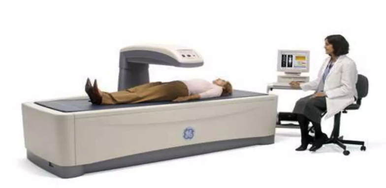
- A DXA scan is the only reliable way to identify reduced bone density before a fracture occurs.
- With a structured, reversal-first clinical approach, measurable improvements in bone density are achievable for women diagnosed with early-onset osteopenia.
- Dietary and lifestyle changes support bone health — but they work best when combined with clinical assessment and monitoring.
At the London Osteoporosis Clinic, we frequently see women in their 30s and 40s who are surprised by a similar diagnosis. The assumption that osteoporosis and osteopenia are conditions of old age is one of the most persistent misconceptions in bone health — and one of the reasons why early bone loss goes undetected for so long. Here is what you should know about early-onset bone loss, why it happens, and what a structured clinical response looks like.
The diagnosis: osteopenia at 37
Paltrow has described undergoing a bone density scan following a tibial plateau fracture — a break at the upper end of the shin bone. The results, she has said publicly, showed bone density consistent with a post-menopausal woman, despite her being in her late 30s. Her doctors identified severely low vitamin D levels as a significant contributing factor.
This pattern — a fracture prompting a scan that reveals pre-existing bone loss — is one we recognise in clinic. Many patients do not know their bone density is reduced until an injury forces the question. It is why we advocate for earlier assessment in anyone with known risk factors, rather than waiting for a fracture to present the evidence.
Why bone loss can start in your 30s and 40s
Bone density peaks in the late 20s and then begins a slow, gradual decline. For most people, that decline is modest and manageable. For some, however, a combination of factors accelerates it significantly — producing the kind of bone density in a 37-year-old that one might expect decades later. The four most common drivers we see in younger patients are:
Low body weight and a history of intensive dieting. Bone responds to mechanical load — the weight the skeleton bears as a person moves through the day. Low body weight reduces this load, and the bone responds accordingly by maintaining less tissue. Additionally, very low-calorie diets or periods of nutritional restriction reduce the availability of calcium, protein, and other nutrients essential to bone matrix. Women with a history of disordered eating, or who have maintained very low weight over time, carry a meaningfully elevated risk.
Vitamin D deficiency. Vitamin D is the primary regulator of calcium absorption in the gut. Without adequate levels, the body cannot absorb dietary calcium efficiently — and it compensates by drawing calcium from bone. Deficiency is common across the UK population, but particularly in those who avoid sun exposure, follow restrictive diets, or have darker skin pigmentation. It is also correctable, which is why we test it at every initial consultation rather than assuming adequacy.
Hormonal changes and early menopause. Oestrogen is protective for bone. It suppresses osteoclast activity — the cells that break down bone tissue — and supports the balance of remodelling. When oestrogen levels fall, whether through natural menopause, surgical menopause, or conditions that affect ovarian function, bone loss accelerates. Women who experience early menopause (before age 45) or who have irregular menstrual cycles over extended periods carry a higher risk. Paltrow has spoken publicly about entering perimenopause in her 40s, which would compound the effects of her earlier diagnosis.
Family history. Bone density has a significant heritable component. A mother or close female relative with osteoporosis or a history of fragility fracture meaningfully increases an individual’s risk. It does not make bone loss inevitable, but it shifts the threshold at which earlier assessment is warranted.
The role of vitamin D — and why it is not optional
Paltrow’s doctors prescribed high-dose vitamin D following her diagnosis — a standard clinical response to severe deficiency. This is not a “wellness” intervention; it is a correction of a biochemical deficiency that has a direct, measurable effect on bone metabolism.
We assess 25-hydroxyvitamin D levels at every initial consultation. Our target for patients in active bone health management is typically 75–100 nmol/L — above the level often described as “sufficient” in general population guidance, and calibrated to what bone metabolism actually requires. Supplementation doses are adjusted to the individual’s baseline, not prescribed uniformly. Taking vitamin D without measuring the starting level risks either under-treating a deficiency or, at very high doses, toxicity.
What an early diagnosis makes possible
Osteopenia is not osteoporosis. It is a signal — an opportunity to intervene before bone loss reaches the threshold at which fracture risk becomes clinically significant. The window between a diagnosis of osteopenia and the development of osteoporosis is precisely where clinical intervention is most effective.
A structured response includes: correcting nutritional deficiencies (vitamin D, calcium, protein); establishing a loading exercise programme that combines weight-bearing and resistance training; assessing hormonal status and, where appropriate, discussing hormone replacement therapy; and monitoring change over time using repeat DXA scanning. Diet and lifestyle are part of this — but the clinical value comes from assessing them individually, not from applying a generic protocol.
Women who receive an osteopenia diagnosis in their 30s or 40s have a meaningful advantage over those diagnosed later: they have time, and they have options. The clinical trajectory is not fixed.
What the evidence shows about early intervention
While we cannot comment on individual cases, our clinical experience with the BoneRevive® programme shows that with a reversal-first approach, measurable improvements in bone density are achievable for women diagnosed with early-onset osteopenia. This approach prioritises rebuilding bone through nutritional optimisation, structured loading exercise, hormonal assessment, and targeted supplementation — with bisphosphonate medication reserved for patients where the risk profile requires it, rather than as a first-line response to a low DXA score.
The principle is straightforward: bone is responsive tissue. Given the right signals — mechanical load, adequate nutritional building blocks, appropriate hormonal environment — it can increase in density. The earlier that programme begins, the more bone there is to work with. Explore our care pathways to understand how we approach this clinically.
Paltrow’s story is useful not because of who she is, but because of what her diagnosis illustrates: that bone loss is silent, that it can begin well before midlife, and that the moment of diagnosis — when caught early — is an opportunity rather than a conclusion. At the London Osteoporosis Clinic, we see this regularly. Patients who are health-conscious, active, and surprised. The assessment answers the question their lifestyle could not. If you have risk factors for early bone loss — a family history, a history of low weight or amenorrhoea, a previous fracture, or simply a concern — a DXA scan with a consultant review is the appropriate next step. We can advise from there.
Frequently asked questions
What is the difference between osteopenia and osteoporosis?
Osteopenia refers to bone density that is lower than average for age but not yet at the threshold classified as osteoporosis. It is measured using a DXA scan and expressed as a T-score. A T-score between −1.0 and −2.5 indicates osteopenia; below −2.5 indicates osteoporosis. Osteopenia does not inevitably progress to osteoporosis — with appropriate intervention, bone density can stabilise or improve.
Should women under 40 be concerned about their bone density?
Most women under 40 do not require routine DXA scanning. However, certain risk factors warrant earlier assessment: a family history of osteoporosis or fragility fracture, a history of amenorrhoea or low body weight, long-term corticosteroid use, a previous fracture, or conditions affecting absorption such as coeliac disease. If you are unsure whether you have risk factors, a consultation with a bone health specialist is a reasonable starting point rather than waiting until a standard screening age.
Can osteopenia be reversed?
Bone density can improve with appropriate intervention, particularly when osteopenia is identified early and the contributing factors — nutritional deficiencies, low mechanical load, hormonal changes — are addressed directly. The degree of improvement varies between individuals and depends on the baseline density, age, and the consistency of the programme. Our clinical experience with the BoneRevive® reversal-first approach shows that measurable improvement is achievable for many patients with early-onset osteopenia. A consultation will establish what is realistic in your individual case.
Medically reviewed by Dr. Taher Mahmud, Consultant Rheumatologist and Co-Founder, London Osteoporosis Clinic. Dr. Mahmud has over 25 years of clinical experience in bone health and osteoporosis management.
This article references publicly available information about Gwyneth Paltrow’s health for educational purposes only. The London Osteoporosis Clinic has no affiliation with Ms Paltrow and cannot comment on her individual medical care. This article does not constitute medical advice. Always consult a qualified clinician for personalised guidance.