Introduction
You’ve likely heard people call MRI scans “safe because there’s no radiation.” But recently, conversations online have raised new questions — can strong magnetic fields in MRI scanners actually affect our DNA? And what happens if you undergo a whole-body MRI every couple of years for health monitoring?
At London Osteoporosis Clinic, we’re passionate about providing evidence-based clarity for patients navigating preventive health choices. This article explores what decades of research say about MRI’s biological safety, what small studies have found regarding potential cellular effects, and why a 1.5–3 Tesla whole-body MRI every two years remains within globally recognised safety standards.
If you’re considering a whole-body MRI as part of a YouOptimised™ or BoneRevive® health assessment, this guide explains what’s really happening inside your body during the scan — and why, when used responsibly, MRI is one of the safest diagnostic tools in modern medicine.
Does MRI Have Any Impact on Health? (Research Overview)
1. Single Routine MRI: No Detectable DNA Damage
Clinical research consistently shows no measurable increase in DNA or chromosomal damage markers after a single, standard MRI.
- Studies reviewed by Stanford Medicine and PMC found no genotoxic effects at diagnostic field strengths (1.5–3 Tesla).
- If any effects exist, they are biologically tiny and within natural cellular variability.
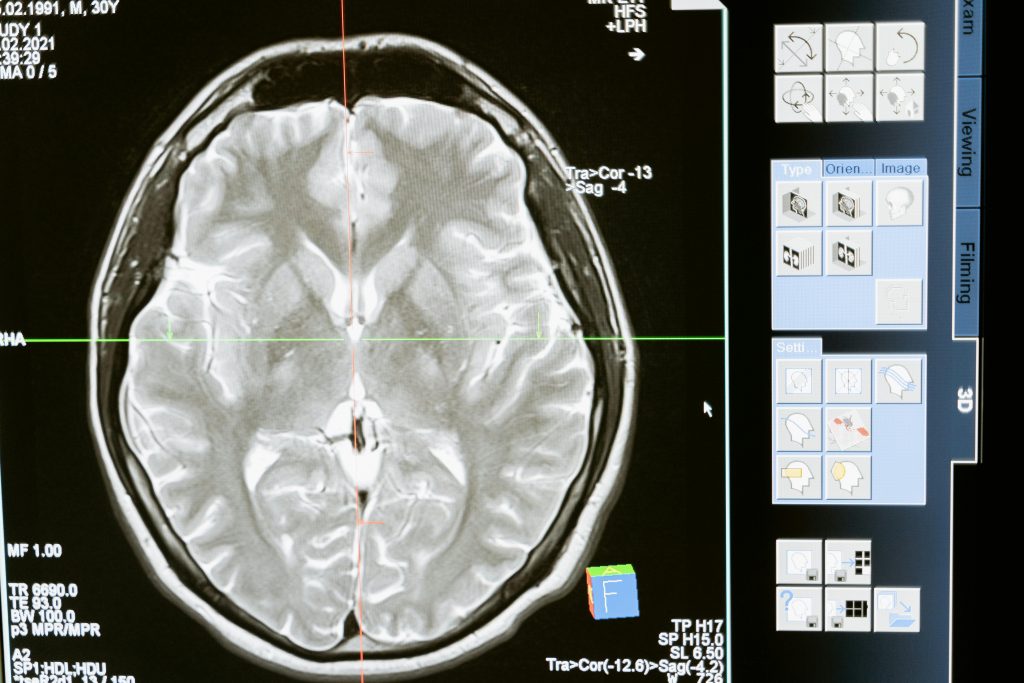
2. Repeated MRIs in a Short Period: Minimal Cellular Changes
A detailed 2022 PubMed study examined individuals who had undergone 10–20 brain MRIs. It detected small, statistically significant increases in some chromosomal break markers — but these changes plateaued and showed no link to illness or disease.
Such findings suggest that the body’s natural repair mechanisms easily manage transient oxidative stress.
3. Ultra-High Field (7 Tesla) Research Context
Experimental studies at 7 T (much higher than routine clinical settings) have observed modest increases in markers like DNA double-strand breaks or micronuclei.
However, these are inconsistent across laboratories, transient, and not associated with actual tissue or organ harm. Routine clinical MRI remains at 1.5–3 T.
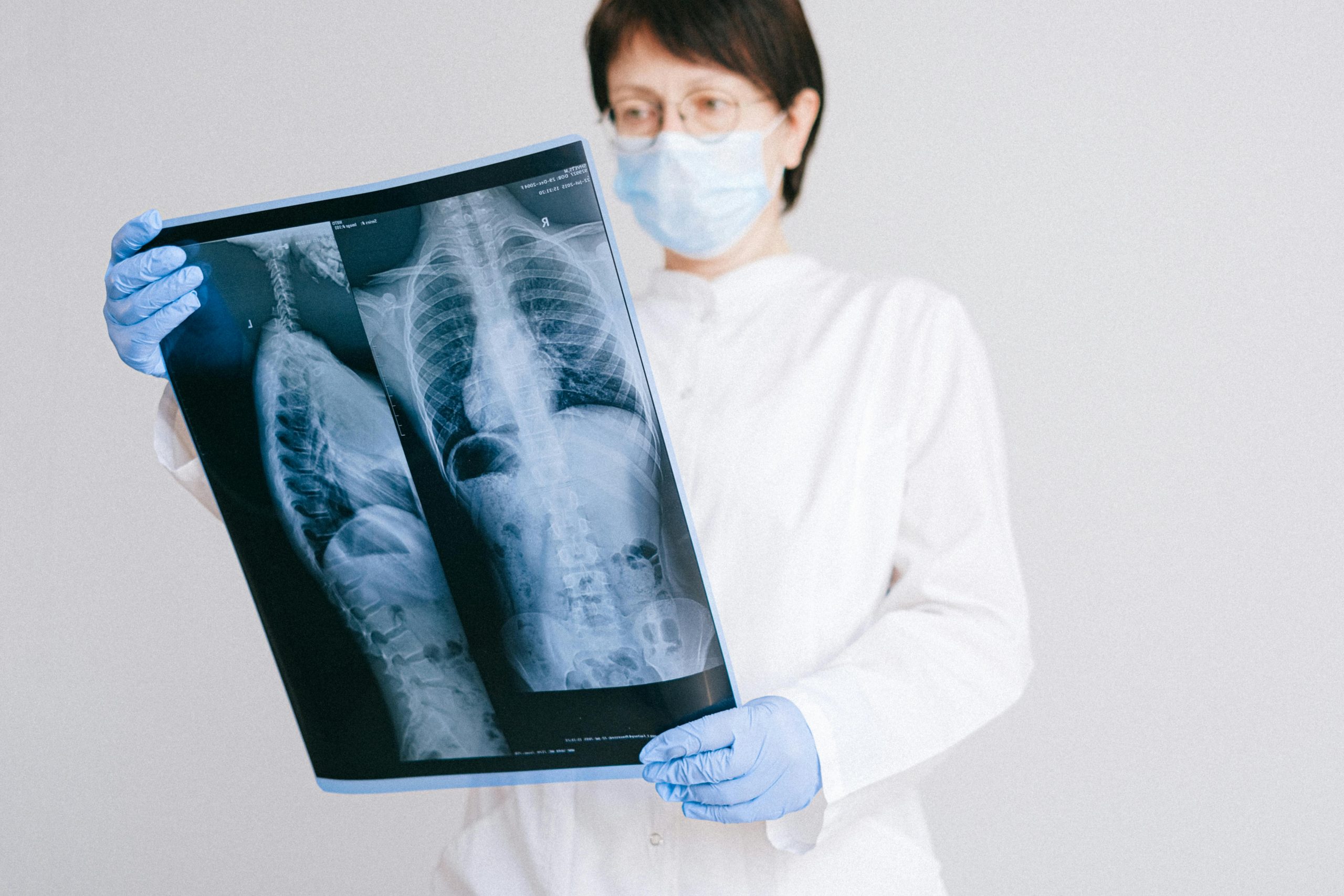
4. Why MRI Is Different from X-rays and CT
MRI uses non-ionising radiation — radiofrequency (RF) and static magnetic fields — unlike X-rays or CT scans, which use ionising radiation that can directly damage DNA.
MRI fields rotate hydrogen nuclei in water molecules; they do not break chemical bonds. This distinction explains its excellent safety record over four decades.
5. System Safety and Exposure Limits
MRI machines are engineered to meet strict IEC, FDA, and MHRA standards.
- Whole-body specific absorption rate (SAR) is limited to 2 W/kg in normal operating mode.
- Resulting tissue heating is typically < 0.5 °C and temporary.
- No credible evidence links MRI exposure to cancer or long-term biological damage (Radiopaedia, ResearchGate).
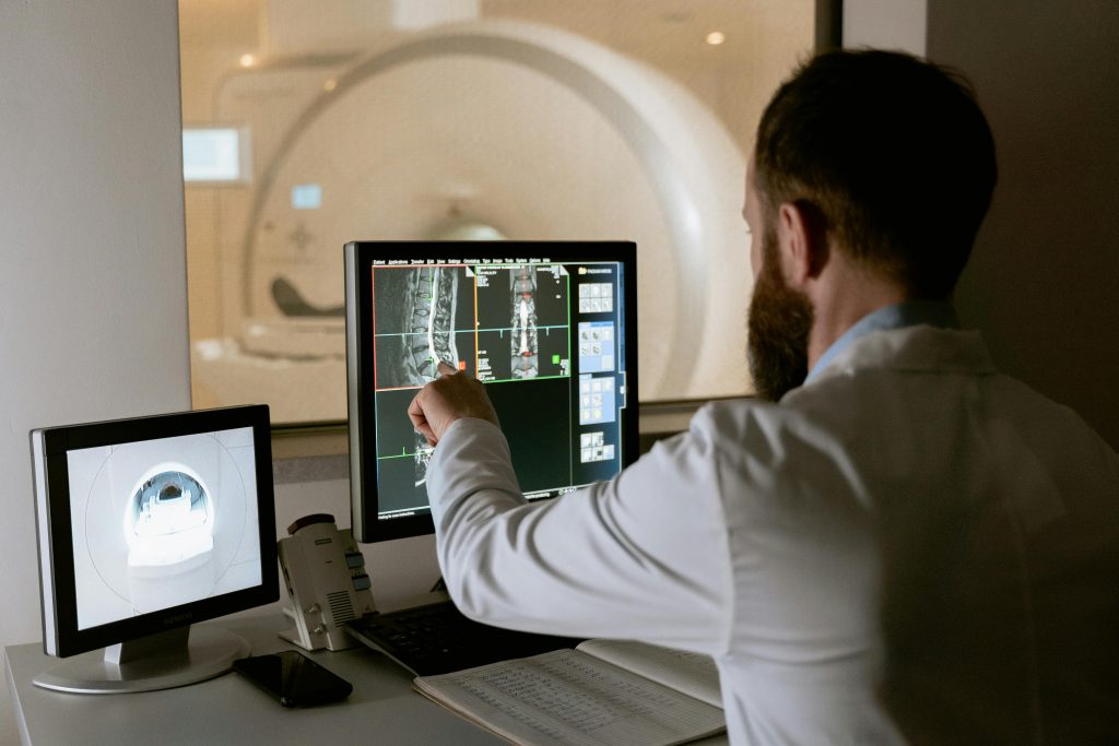
Interpreting the Evidence Clinically
In everyday radiology practice, there is no evidence that MRI causes genetic or cellular harm at diagnostic levels.
The few studies noting small cellular changes rely on surrogate blood markers, not disease outcomes. Moreover, these markers fluctuate naturally in healthy individuals.
For patients requiring multiple MRIs (for example, neurological surveillance or oncology follow-up), benefits far outweigh theoretical risks. As with any procedure, clinical necessity and optimisation remain key principles.
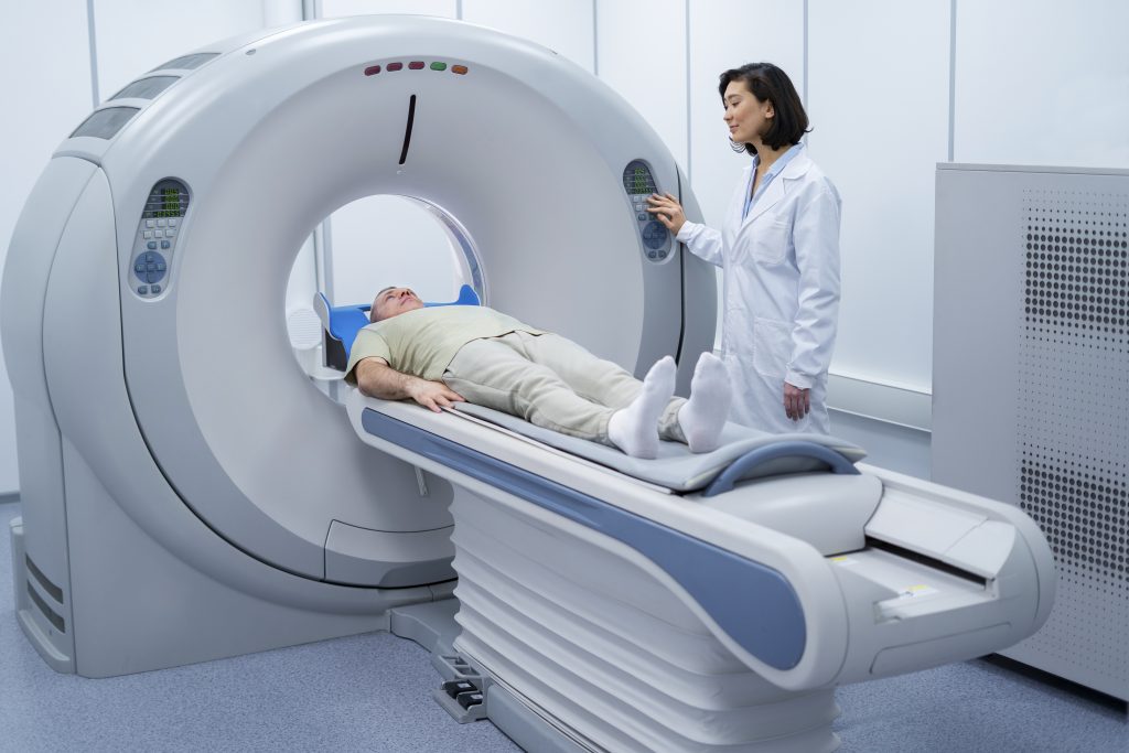
Is Whole-Body MRI Every Two Years Safe?
For preventive health screening or ongoing wellness monitoring, many patients now consider whole-body MRI (WB-MRI) every one to two years. The evidence remains clear and reassuring.
1. Scientific and Clinical Context
- Field Strength: Standard WB-MRI uses 1.5–3 T — identical to routine brain or spine MRIs.
- Energy Type: The scan involves static magnetic fields and brief RF pulses, both non-ionising.
- Thermal Effect: SAR-controlled heating results in < 0.5 °C tissue rise — transient and safe.
2. DNA and Cellular Integrity
Across decades of human and animal studies, no consistent evidence shows DNA strand breaks, chromosomal abnormalities, or mutations following routine MRI.
Even the small, transient changes seen in intensive-exposure research fall within natural biological variation and are easily repaired by normal cellular processes.
3. Cumulative and Long-Term Effects
There is no known cumulative damage mechanism for MRI’s non-ionising fields.
Even weekly MRIs in research or clinical surveillance have not demonstrated adverse outcomes.
At a two-year interval, exposure is far below all international safety thresholds.
Global Health Authority Positions
- World Health Organization (WHO): MRI “has not been shown to have any deleterious health effects when used within recommended guidelines.”
- FDA / IEC / MHRA: Approve routine diagnostic MRI up to 3 T for all populations, including children and pregnant women (post-first trimester).
- European Society of Radiology: Considers WB-MRI safe and suitable for specific screening and preventive contexts.
Potential Non-Biological Risks
While biological risk is negligible, a few practical aspects deserve consideration:
| Category | Issue | Comment |
|---|---|---|
| Claustrophobia / anxiety | Common, manageable with reassurance or mild sedation | |
| Gadolinium contrast | Rare allergic or retention concerns (not relevant for non-contrast scans) | |
| Incidental findings | May trigger unnecessary follow-up | Experienced radiologist interpretation recommended |
| Metal implants | MRI-unsafe implants may contraindicate scanning | Screening essential |
MRI in Bone and Systemic Health Monitoring
At London Osteoporosis Clinic, whole-body MRI can play a meaningful role in preventive care.
It provides a radiation-free view of bone structure, muscle mass, marrow health, visceral fat, and vascular status — aligning with comprehensive health assessments such as:
- BoneRevive®: Focused on bone strength and fracture prevention.
- YouOptimised™ Health MOTs: Targeted towards holistic, preventive monitoring.
By identifying subtle musculoskeletal and metabolic changes early, clinicians can personalise interventions long before disease develops.
Summary: The Evidence at a Glance
| Factor | Evidence / Consensus |
|---|---|
| Magnetic field 1.5–3 T | Safe; used globally for >40 years |
| Frequency | Non-ionising; no DNA bond breakage |
| Interval | Every two years ≪ safety thresholds |
| Cumulative risk | None demonstrated |
| Main concerns | Claustrophobia, contrast allergy, incidental findings |
Conclusion and Takeaway
So, would an MRI have any impact on your health?
According to decades of scientific data and global safety standards, the answer is no — not in any clinically meaningful way. MRI is non-ionising, non-destructive, and engineered within conservative safety margins. Even among patients undergoing multiple scans, no pattern of illness, mutation, or cancer has emerged.
A whole-body MRI once every two years comfortably fits within all recognised exposure limits. The potential “risks” are logistical — cost, comfort, and the possibility of false alarms — rather than biological.
At London Osteoporosis Clinic, we view MRI as a valuable ally in preventive medicine: a non-invasive, evidence-based tool for tracking musculoskeletal and systemic health safely over time.
If you’d like to explore how whole-body MRI could integrate into your personalised health plan, visit our https://www.londonosteoporosisclinic.com page or contact the clinic to discuss a tailored assessment.